What is Computed Tomography?
 Computed Tomography (CT) provides a welfare friendly way of assessing the total muscle, fat and bone yield in a live sheep using whole body X-ray image analysis. This near perfect predictor of carcase composition can also be used to measure the muscling in different parts of the carcase, such as the gigot.
CT is a successful tool for identifying outstanding animals within the breed, but it has also had an equally important wider impact on breeding improvement.
The use of CT has enhanced our understanding of the relationship between on-farm ultrasonic measurements and lean and fat in the carcase. This has improved the efficiency with which superior animals can be identified using on-farm ultrasound and strengthens the breeding evaluations produced across the breed. CT scanning is a great tool to identify superior genetics within terminal sire flocks and can assist the marketing of recorded rams.
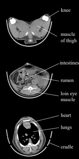
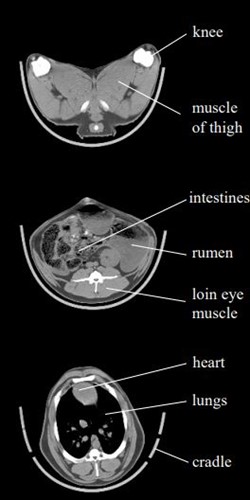
Diagram 1. Three CT images
Positions of the three cross-sectional scans on the right are shown on the lefthand scout scan.
Key anatomical features are labelled on each diagram for orientation.
NB: CT measures density. The greyscale used for the cross-sectional scans shows air as black, fat as dark grey, muscle as light grey and bone as white. The sheep is lying on its back in the cradle.
The economics of using CT
It will never be economically or logistically possible to scan all potential breeding animals. In the UK a two-tier approach is used to identify candidates for CT scanning. All lambs scanned using ultrasound and the best are then sent to the CT scanner.
What will CT scanning tell you?
Raw CT measurements will show the:-
- Weight for fat, muscle and bone in the carcase
- Percentage of fat, muscle and bone in the carcass
- Killing out percentage (total tissue weight / liveweight)
- Ratio of muscle to bone and muscle to fat in the carcass
- Distribution of muscle in the carcase – including the percentage of muscle in the leg, loin or chest
- Gigot shape
The accuracy of Estimated Breeding Values for growth and carcase traits will be greatly enhanced through the incorporation of CT data into the evaluation of your flock. Indexes and EBVs will be increased if CT measures prove an individual or family to be genetically superior.
What does CT scanning cost?
The amount that SRUC charges for CT scanning varies depending on whether breeders used the static CT scanner in Edinburgh or the mobile CT service. Levy body subsidy is often available from AHDB, HCC and QMS.
Subsidy is subject the following conditions:
- Lambs are male and scanned using ultrasound at an acceptable age.
- At least five ram lambs must be sent per participating farm.
- Lambs must have been reared under similar conditions.
- Lambs must have an individual identification that can be readily linked to information on the Signet database.
What do I do next?
Contact Kirsty McLean at SRUC to get your lambs booked in.
- Contact details for Kirsty are Tel: 0131 535 3250 / 3251 Fax:0131-535-3404
Email: [email protected] / Website - Let your ultrasound scanning technician know that you wish to CT scan, so that an appropriate date can be set for ultrasound scanning.
- Talk to your Breed Society to see if it is possible to share transport with other breeders when taking lambs to Edinburgh.